Tumor

Tumor head / brain

Head tumors comprise those of the brain and those of adjoining regions, namely meninx (cerebral membrane), bones, skull base, petrous bone, paranasal sinus, and eyes.

Tumors are classified into benign and malignant tumors (cancer).

Within the group of intracranial (inside the head) benign tumors, especially the so-called cerebellopontine angle tumor (acoustic neurinoma) and those of the meninx (meningeom) occur most frequently. They can be completely removed by microsurgery and, thus, are curable.

Within the group of malignant tumors, metastases are most frequently. The origin of metastases is parts from a primary tumor (e.g., lung or intestines) that have circulated through vessels and deposited in normal tissue. Primary tumors describe the tissue from which metastases orignate.

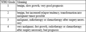
Besides abovementioned classifications, there are malignant primary tumors, the so-called glioma and astrocytoma. These are graded in terms of their malignancy: grade II to IV. Grade IV corresponds to the most severe and aggressive tumor, the glioblastoma. Healing is only possible in very few cases. The function of neuroradiology and neurosurgery here is to ensure the correct diagnosis (by differentiating it from diseases with similar symptoms which are potentially curable), to preserve quality of life and prolong life in general.

Besides the tumor grading, there is also a classification of the tumors of the central nervous system according to the WHO. You can watch or download it here.

Symptoms brain tumor

The symptoms of brain tumors highly depend on their location, their growing pace and their size. The following examplary symptoms can occur:

  • headache
  • epileptic fits
  • character changes
  • absent-mindedness
  • tiredness, loss of power
  • cerebral focal disorders, for example
    • paralyzation of one body side (hemiplegia)
    • paralyzation of cerebral nerves
    • aphasia (language disorder)
    • apraxia (motor disorder)
    • agnosia (loss of ability to recognize objects, for instance, proper visual perception but inability to recognize faces)
    • amnesia

Diagnostics brain tumor

Tumor diagnostics include imaging examination – primary state of the art is MRI (magnetic resonance tomography) – and neurosurgical biopsy, i.e., taking tissue specimen.

Besides the depiction of anatomic structures by means of contrast agents or without them, MRI makes it possible to examine metabolic processes, for instance, by employing diffusion or perfusion imaging, or spectroscopy. Since these examination techniques analyze functional processes of the body, they are often called functional magnetic resonance imaging (fMRI). Presurgically it is possible to distinguish tumor regions from adjoining functional areas and nerve tracts by using fMRI BOLD imaging and tractography (diffusion tensor imaging, DTI).

A CCT (cranial computed tomography) is primarily conducted to plan surgery, or if calcification or other issues have to be clarified which necessitate computed tomography (CT) besides MRI.

Treatment brain tumor

Dependent on degree of severity, benignancy/malignancy, the tumor is to be removed completely, if possible. Today of microsurgery is the method of choice, during which the surgeon is able to navigate to the affected tissue accurate to the millimeter. If the tumor cannot be removed completely, additional radiation or chemotherapy is to be applied.